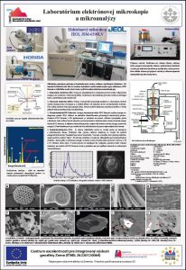
SEM (scanning electron microscopy) laboratory is equipped with JEOL JSM-6390LV electron microscope. The sample is bombarded with an electron beam. The electrons interact with atoms in the sample, producing various signals that can be detected and that contain information about the sample's surface topography and composition. Specimens can be observed in high vacuum or in low vacuum. The types of signals produced by an SEM include secondary electrons (SE), back-scattered electrons (BSE), characteristic X-rays, light (cathodoluminescence) (CL).
For SE imaging, the electron microprobe functions as an SEM, providing topographic information about a sample. The spatial resolution for SE imaging is depending on the accelerating voltage, beam current, and other operating conditions. Common applications include studies of grain morphology, precipitates on a mineral surface, microfossils (for instance, ancient diatoms), and other materials too small for visible light microscopy.
BSE images show atomic number differences within a sample. A certain fraction of the electrons in the beam are scattered "backward" out of the sample as a result of interactions with its nuclei, and these "backscattered" electrons can be used to form images. The number of electrons are backscattered increases with increasing mean atomic number of the material. In a BSE image, the brighter the area, the heavier the mean atomic mass of that material.
Characteristic X-rays that are produced by the interaction of electrons with the sample may also be detected in an SEM equipped for energy-dispersive X-ray spectroscopy (OXFORD Instruments INCA x-act). Analysis of the x-ray signals may be used to map the distribution and estimate the abundance of elements in the sample.
Cathodoluminescence, the emission of light when atoms excited by high-energy electrons return to their ground state, is analogous to UV-induced fluorescence. Our microscope is equipped with HORIBA FCLUE/MHRA with two detectors PMT and CCD (UV-Vis-NIR). In the SEM, CL detectors either collect all light emitted by the specimen or can analyse the wavelengths emitted by the specimen and display an emission spectrum or an image of the distribution of cathodoluminescence emitted by the specimen in real color.
Contact person:
+421 48 321 3312
Workplace:
Banská Bystrica, Ďumbierska 1, Geological Division